At 14, Xiao Zhang should have been enjoying her school life to the fullest. Instead, she was plagued by recurrent abdominal pain that grew increasingly severe over the past two years. What began as occasional, brief episodes progressed to frequent bouts—sometimes weekly or even daily—with pain lasting hours instead of minutes. The discomfort shifted locations unpredictably, disrupting her studies and daily life.
Despite seeking medical help earlier, blood tests showed no abnormalities. Then, just before an important exam, another intense and prolonged pain attack sent Xiao Zhang to the hospital for an abdominal ultrasound—this time revealing multiple suspicious foreign objects in her small intestine. Baffled, she couldn’t recall swallowing anything unusual. When local doctors couldn’t pinpoint the cause, they referred her to Renji Hospital in Shanghai for further evaluation.
At Renji’s Department of Gastroenterology, doctors promptly arranged a small bowel CT and capsule endoscopy—non-invasive imaging that provided a comprehensive view of her intestines. The results confirmed over a dozen hollow, pebble-sized objects in her pelvic small intestine, clearly the source of her pain. While surgical exploration might have been necessary to identify and remove them, Renji’s team leveraged their expertise in double-balloon enteroscopy (DBE), a minimally invasive technique the hospital pioneered in China. With vast experience in small bowel interventions, they admitted Xiao Zhang and scheduled the procedure.
Before the DBE, doctors conducted thorough evaluations of her nutrition and medical history, with anesthesiologists ensuring safe sedation. During the procedure, Dr. Xie from Renji’s endoscopy team identified pale yellow, stone-like objects (each 2–3 cm wide) obstructing the jejunum. Using precise tools, Dr. Xie carefully extracted the objects.
But the mystery deepened: Xiao Zhang had no history of pica or ingestion of foreign bodies. When lab analysis revealed the "stones" were composed solely of bile acids—with no external materials—Dr. Xie suspected a congenital intestinal malformation trapping digestive residues. To prevent recurrence, surgery was needed to remove the affected segment. Gastrointestinal surgeons successfully excised a large congenital jejunal diverticulum, the root cause where food and fluids had stagnated, forming the stones.
Xiao Zhang was discharged a week later. A year on, her abdominal pain hasn’t returned. The small intestine’s length and complexity often make diagnoses challenging. For rare cases like Xiao Zhang’s, Renji Hospital’s combination of clinical expertise, cutting-edge enteroscopy, and multidisciplinary collaboration ensured accurate detection and effective treatment—turning a perplexing condition into a resolved chapter of her life.
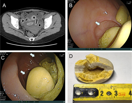
Figure: CT/DBE results and the extracted stones. White arrows indicates the diverticulum.